- PDF (5 MB)
- Pages document (8 MB)
- iBook (10.5 MB)
- iBook file (22.8 MB) for iAuthor
- 27 8.5×11 pages with a cover, introduction, table of contents and answers
- March 2010: Minor changes to delta gap
- June 2011: Fixed error in Metabolic alkalosis (thanks Rakesh Lattupalli) and some spelling errors
- September 2011: fixed three typos, reworked anion gap, non-anion gap, and rapid interpretation of ABG introduction
- December 2011: fleshed out osmolar gap, smoothed the introduction to primary acid-base disorders. Typos: some removed and some added.
- January 2012 iBook!
- March 2012 added a picture and graph to osmolar gap. Moved the files to drobox. Added the iAuthor file for download.
- Jan 2013 Major revision. New cover, new intro, added two additional clinical vignettes, 2-pages on DKA, 2 pages on RTA including urine anion gap.
OUWB Question: Acid-Base
Hi Dr. Topf,
(I don’t have Twitter) I wanted to ask you about question 6 on the week 2 quiz:
“An unresponsive woman is brought to the emergency room. She has a history of a suicide attempt a few years earlier. The lab tests are: Serum Na 140 mmol/L Serum K 4.0 mmol/L Serum Cl 100 mmol/L Serum HCO3 14 mmol/L, BUN 17 mg/dl, creatinine 0.7 mg/dL, serum osmolality 323 mOsm/Kg, Blood glucose 72 mg/dl, Blood gases: pH 7.28 pCO2 27 mmHg. What would you expect the urine pH to be in this patient?”
Why is it that we would expect the urine pH to be acidic? Since blood pH is 7.28, I would imagine that urinating out HCO3- (explaining the low serum HCO3) would have caused the acidic blood pH, thus making urine pH basic?
Thanks for your help,
“An unresponsive woman is brought to the emergency room. She has a history of a suicide attempt a few years earlier.
This is the “tell” of the stem. Acid base + suicide = ethylene glycol toxicity
The lab tests are: Serum Na 140 mmol/L Serum K 4.0 mmol/L Serum Cl 100 mmol/L Serum HCO3 14 mmol/L, BUN 17 mg/dl, creatinine 0.7 mg/dL , Blood glucose 72 mg/dl,
They don’t tell you the anion gap. Calculate it.
High anion gap.
serum osmolality 323 mOsm/Kg
More of the tell. They won’t tell you the osmolality unless they want you to calculate the osmolar gap (or it is a hyponatremia question)
Osmolar gap = 323 – (280 + 4 + 6 + 0)
Osmolar gap = 323 – 290
Osmolar gap is a massive 33 (Upper limit of normal is 10, over 20 starts to gain a lot specificity for toxic alcohol)
This confirms our earlier suspicions of ethylene glycol toxicity
Blood gases: pH 7.28 pCO2 27 mmHg. What would you expect the urine pH to be in this patient?”
The ABG confirms the metabolic acidosis.
Let’s do Winters formula (not really needed for this question, but you know…practice)
1.5 x 14 =21 + 8 =29, measured CO2 is within ±2 of predicted so an appropriately compensated metabolic acidosis.
Why is it that we would expect the urine pH to be acidic? Since blood pH is 7.28, I would imagine that urinating out HCO3- (explaining the low serum HCO3) would have caused the acidic blood pH, thus making urine pH basic?
So the bicarbonaturia you are talking about would happen if the cause of the metabolic acidosis is renal loss of bicarbonate (what we call renal tubular acidosis).
The urine would also be acidic if the patient had a non-anion gap metabolic acidosis from diarrhea.
Hope this helps
hypokalemia and metabolic alkalosis
A few years ago I was talking one of my mentors at Kidney Week, John Asplin. He mentioned
that he taught an integrated lecture on metabolic alkalosis and hypokalemia. I thought this was an inspired idea.
Teaching separate classes on both subjects results in a lot of overlap because the renal mechanisms for both disease are the same, this means that many of the diseases that cause one, also cause the other.
Additionally hypokalemia can cause metabolic alkalosis and metabolic alkalosis can cause hypokalemia, so it makes sense to teach both of these conditions in an integrated lecture.
Lastly, teaching each electrolyte individually in isolation from each other is a missed opportunity. One can only appreciate the beauty of electrolyte physiology when one understands how each electrolyte fits together and how abnormalities in one is associated and affects all of the other electrolytes.
Unfortunately, I botched the lecture. I gave this lecture for the first time for the Oakland University Beaumont Medical School this past August. I knew it didn’t go too well, but this week I received the class feedback. Overall my statistical evaluations were excellent but when I read the comments the students were jackals. They savaged this lecture.
Timing was on my side, I was scheduled to give this lecture the day after I received feedback. I’m not done tweaking it but what I did for my Tuesday lecture was add more connective tissue between the concepts, and fill in with some additional summary slides.
Right now, I’m using it as a lecture to follow-up my potassium lecture, but at OU the students didn’t have any baseline potassium knowledge. In order for this lecture to work the students must already understand the basics of potassium, especially the central role that renal potassium handling has in potassium homeostasis. Hopefully I will be able to negotiate another hour into the GU schedule for this lecture.
My next plans for this lecture is to cut out a lot of the opening slides. The purpose of those slides is to quickly move from introducing potassium and hypokalemia to getting to the truth that hypokalemia is almost solely a disease of increased renal losses.
I want to add a slide about disease opposites:
- Pseodohypoaldosteronism type 1 and Liddle syndrome
- Godon’s syndrome and gittleman’s syndrome
- Adrenal insufficiency and AME
I want to add some slides on how hypokalemia causes (specifically, maintanes) metabolic alkalosis and then how metabolic alkalosis causes hypokalemia.
Non-anion gap metabolic acidosis screen cast
Altitude sickness and the role of acetazolamide
I am going to Kings Canyon National Park at the end of the month. I will leave Detroit, elevation 600 feet and will travel via planes, trains and automobiles to 9,000 feet for the first night. Then we will begin out hike and cross passes over 12,500 feet.
In the past, I have developed modest altitude sickness going from 600 to 8,000 feet. So, I am nervous about the same problem on this trip. Acetazolamide is supposed to ameliorate altitude sickness.
The body acclimates to decreased oxygen and is so effective that people can function at the top of Mt. Everest without supplemental oxygen. The partial pressure of oxygen at the summit is 43 mmHg which is equivalent to breathing 6% FiO2.
From NEJM 2009, 360: 140-9 |
The primary means of improving oxygenation is hyperventilation. Hypoxia stimulates ventilation. There is also an increased ventilatory response to carbon dioxide so that that the normal respiratory response to carbon dioxide is exaggerated so that one gets more ventilation at lower CO2 levels. The reason that increased ventilation improves oxygenation has to do with the effect carbon dioxide in the blood has on oxygen transfer in the alveoli. During respiration CO2 leaving the blood dilutes the incoming oxygen at the alveoli, increased respiration, lowers the pCO2 and hence minimizes this dilution.
Antagonizing the hyperventilatory response is respiratory alkalosis. Central chemoreceptors detect alkalosis in the CSF and slow respiration. This is one of the key factors preventing the essential hyperventilation.
Acetazolamide (Diamox) is a carbonic anhydrase inhibitor. Carbonic anhydrase catalyzes the reaction converting bicarbonate to carbon diaoxide and water:
This is the fundamental buffer reaction in the body and it is amazing to me that blocking this essential acid-base reaction is not lethal. Acetazolamide works in the proximal tubule by blocking the reabsorption of filtered bicarbonate.
Acetazolamide induces a proximal renal tubular acidosis (RTA 2). This results in metabolic acidosis. The metabolic acidosis stimulates compensatory hyperventilation. This metabolic acidosis antagonizes the respiratory alkalosis which normally occurs with hyperventilation.
Their maybe additional advantages of acetazolamide including decreased CSF production and antagonizing fluid retention.
Happy climbing.
Abacavir and methanol poisoning
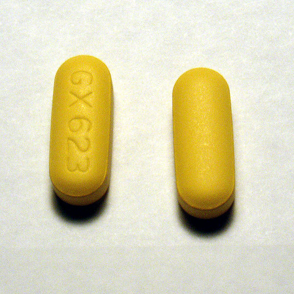
About a month ago, Nephron Power wrote about a great electrolyte case in AJKD. The case regarded a patient who drank a liter of methanol but was asymptomatic. The reason the patient was apparently resistant to a toxic methanol slug of methanol (The quantity of methanol that produces toxicity ranges from 15 to 500 ml of a 40% solution to 60 to 600 ml of pure methanol) was protective powers of abacavir. Abacavir is a nucleoside reverse transcriptase inhibitor and apparently, is a potant inhibitor of alcohol dehydrogenase, the critical enzyme which converts methanol into formaldehyde. Formaldehyde is then converted into the lethal formic acid by formaldehyde dehydrogenase.
After reading this I started to wonder if abacavir was such an effective inhibitor of alcohol dehydrogenase what happens when patients get exposed to say a more common substrate of alcohol dehydrogenase such as whiskey. Shouldn’t we hear about people on abacavir going on alcohol benders after a single shot of ethanol?
A couple of cracks at PubMed and I sure didn’t find much. Barber, Marrett et al. looked for two types of alcohol reactions from abacavir, either a disulfaram-like reaction or reduced alcohol tolerance. The authors found three cases of in 173 patients starting abacavir. They found one disulfarem reaction (nausea, tachycardia, flushing with a single shock of vodka) and two cases of decreased alcohol tolerance
After three glasses of wine he felt as though he had a bottle and a half, with memory loss.
The only other paper I could find was by McDowell, Chittick, et al. who looked at increased abacavir levels with alcohol intake. The reverse of what I was looking for, but at least it was related. They gave a single dose of abacavir and 0.7 g/kg of ethyl alcohol to 25 HIV positive men. They found a 26% increase in the half life of abacavir with alcohol but…
This study did not demonstrate any alteration in the pharmacokinetic parameters of ethanol by abacavir coadministration; blood ethanol median profiles following ethanol administration in the presence and absence of abacavir were essentially superimposable. There was no evidence that co-administration of abacavir interferes with ethanol metabolism. There were no disulfiram-type reactions in any subject who received coadministration of abacavir and ethanol.
This study tested the effects of a single dose of abacavir, chronic dosing may result in a different effect on alcohol dehydrogenase.
Interesting case nonetheless.
Acid-Base Chapters (Chapters 10-16) from Fluids
Chapter 10: Introduction to Acid-Base
Delta anion gap. Not as good as we think it is.
One of the concepts that is regularly taught in the evaluation of acid-base status is determining if there are multiple acid base disorders by evaluating the ratio of the delta anion gap/delta bicarbonate.
I teach this concept as determining what the bicarbonae would be in the absence of or prior to the anion gap.
The concept comes from the idea that for every mEq of bicarbonate that is consumed by the strong acid (other anion) the anion gap should rise by one. So if the bicarb is 16, a delta of 8, we would expect an anion gap of 20, a normal anion gap of 12 plus the delta bicarbonate of 8. This is a ∆AG/∆Bicarb of one.
[Some authors] suggested that mixed disturbances should be considered if the ratio is less than 0.8 or greater than 1.2. Paulson, applying this rule to a group of normal control subjects and patients with simple metabolic acidosis, noted that the formula erroneously categorized 56% [specificity of 44%] of this group as mixed disturbances. Use of the 95% confidence interval of ±8 mEq/L increased the specificity to 97% but with a poor sensitivity of only 27%.
RTA Lecture for Medicine Board Review
Introducing the Acid-Base Machine
Last month I realized that in order for students to really learn acid-base interpretation they need practice. Lots of practice. So I started everyday’s teaching rounds by assigning each student four ABGs to interpret.
Creating all of those ABGs became pretty tedious so I started fiddling around with an excel spreadsheet to automate the process. I used ABG Machine 1.0 for that month but, unfortunately, it created too many respiratory problems and too few metabolic disorders. I completely re-crafted the randomization algorithm so that it should provide a balanced distribution of ABG problems (a quarter metabolic acidosis, a quarter metabolic alkalosis, a quarter respiratory alkalosis and a quarter respiratory acidosis).
Introducing The Acid-Base Machine 2.0
The spreadsheet is made up of 12 individual sheets. The first one is the Question sheet, you should print one copy for every student or resident. The second sheet is the Answer sheet, I printed one for me and one for each student and I would pass them out after the excercise so if they wanted to brush up on some additional questions they had the answers. The next ten sheets are the guts of the machine and you can ignore them unless you want to tinker with how the randomization works.