Rise of the Tweetorial

One of the interesting developments in MedTwitter has been the chained tweet to demonstrate a point. I think the master of this is Professor Darrel Francis.

This one is nicely relevant to this week’s NephJC:

Another famous innovator of the Medical Tweetorial is Vinay Prasad. Vinay smartly collects the first tweet to his Tweetorials in a pinned tweet.

https://twitter.com/vinayprasadmd/status/1007337958846783488?s=21

Another master of the Tweetorial is Tony Breu. He, similarly, collected his Tweetorials in one place.

This one has relevance to nephrology. Brilliant explanation.

Speaking of relevance to nephrology, Swapnil has thrown his hat in the Tweetorial ring

Paul Sufka has joined the party focusing with a focus on rheumatology:

Here is Bryan Vartebedian’s take on this development. His problems with the rise of the tweetorial can be summed as:

  • Poor indexing and search means these tweetorials will not be able to be found later.
  • Using 280 character tweets to convey 500-word ideas is a mismatch between the medium and message. These ideas would be better conveyed in a blog post rather than chopped up into beads of text to be strung together.
  • MedTwitter is a just a small sliver of the medical community and doing tweetorials traps the ideas in this small box.

In his conclusion, Vartabedian hedges a bit, but it is clear he believes in the blog:

I firmly believe that all of us should be poking at these applications to discover their most creative uses. But what any of us think is less relevant than what sticks with the public community of physicians. The market will bear this one out.

I love Sufka’s lessons on ANA. Prasay’s thread is a bold call to action that challenges the medical industrial publishing complex. It’s an important reminder that we are the publishers and no longer live in a permission-based system of launching ideas to the world.

We just need to remember that there’s a big medical world and a whole lot of eyeballs beyond our Twitter space. Let’s put our brilliance in the right place.

The new divide

When I was a fellow at University of Chicago, just after the turn of the century, I did literature searches and then divided my PubMed bounty into articles I could download directly and those articles I would need to stalks the stacks to retrieve from hardcover bound versions of the journal. Any article in the latter group needed to be really important for it to be worth the extra-effort.

It was bits versus atoms. Bits were so much easier to gather, manage, and organize that it affected the information I would collect.

This artificial division largely fell away as more and more of the archives were moved to electronic format and the stuff that was trapped in atoms became less relevant as it faded further and further from the current time. Today, effectively all the medical literature exists as bits.

The new barrier is not between bits and atoms but between open access bits and paywalled bits.

See this exchange:

https://twitter.com/akshayakeerti/status/1011754523663261698?s=21

We may be moving to a world where if you want to be read (and to be cited) you will need to publish in open access journals.

Unintended consequences: Oxalate nephropathy and bariatric surgery.

Near the end of Swapnil’s sweeping summaries of the renal denervation articles for this week’s NephJC, he discussed safety. In the trials, the procedure was remarkably safe but Swap brought up possible future consequences. These are adverse events that could never be measured in a real-world trial:

Renal denervation is permanent. Blood pressure medications can be stopped. Why does this matter? Maybe if a patient subsequently develops sepsis or hypovolemia? The sympathetic system exists for a reason….See this case report. In this study of sheep with CKD, bleeding caused a much more severe drop in blood pressure.

This reminds me of bariatric surgery. We had been doing Roux-en-Y for weight loss for decades (See meta-analysis) when, in 2005, Nelson published a case series of calcium oxalate stones, including a couple of cases of ESKD with dialysis dependence due to oxalate nephropathy. Their work was backed-up in this report.

A massive meta-analysis ofover 700(!) studies of weight loss surgery from 1990 through 2002.

Oxalate nephropathy is secondary to increased oxalate absorption following the Roux-en-Y procedure. There are two theories as to why this is:

  1. Fat malabsorption, one of the goals of the therapy leads to saponification of intestinal calcium. Since calcium is unavailable to bind and trap oxalate in the gut more is available for passive and active oxalate absorption in the distal ilium.
  2. Decreased bile acid reabsorption in the gut leads to unconjugated bile salts damaging the luminal membrane and increased passive oxalate absorption.

Additionally the malabsorption leads to diarrhea and metabolic acidosis. This decreases urine pH and lowers urinary citrate both of which promote calcium oxalate stones. There is also a decrease in urine volume of about 0.5 liters, further increasing the stone tendency of these patients. Dr. Park defined a lot of this in a prospective study of 24-urines after Roux-en-Y.

I have seen patients with ESKD from Roux-en-Y. It is horrible. Since the primary pathology has not ceased, these patients are at risk for the same oxalate nephropathy following a transplant, making a transplant a risky procedure.

Though oxalate nephropathy after Roux-en-Y does occur, obesity itself is a risk factor for ESKD and bariatric weight loss appears to protect patients from this.

From Chang et al. Kidney International Reports 2017 2(2): 261-270. (Another good reference on ESKD risk after bariatric surgery)

This good news tempered with the risk of disaster makes the decision to go for bariatric surgery a bit more nuanced than it is often portrayed. Does renal denervation have similar future land mines just waiting to be exploded in the future? We have no idea what unexpected horrors from renal denervation lie undiscovered.

E-mail from my first trip to Kidney Week

My wife and I celebrated out eighteenth anniversary last night. We went to dinner in Windsor. While we were in line waiting to cross back into the US, Cathy found and read some of our e-mails back and forth from the first few years of courtship.
Here is one that I sent from my very first first trip to Kidney Week, which wasn’t even called that back then.
From: Joel Topf
Date: October 25, 1998 at 2:59:00 PM EST
To: Cathy
Subject: I met my hero

Cathy, the conference is awesome. I’m having fun.

I just met Burton Rose. He’s the reason I’m going into nephrology. It was a moving experience.

I’ll call you later
love you
always
j

Hyponatremia fan fiction

I think I may have buried the lead in the last blog post. Perhaps this imaginary conversation will clarify what is so important about the George et al study.

Nephrologist: There are now clinical practice guidelines, based on expert opinion, that tell us to correct sodium no more than 8 mmol/L in the first day of hyponatremia.

Internist: That seems cautious. How well are we meeting those guidelines?

Nephrologist: Well in a recent multi-center, retrospective analysis, of nearly 1500 people with an initial sodium below 120, just over 40% went too fast.

Internist: FORTY PERCENT were too fast. Oh my god! We’re doing horrible!

Nephrologist: Yeah, it’s kind of embarrassing.

Internist: So what happened to the 600+ people where the speed limit was exceeded?

Nephrologist: Well, they found 9 people had osmotic demyelination on MRI. But none of them had any documented permanent neurologic deficits and none of them was diagnosed with central pontine myelinolysis.

Internist: So how many people could we help if we worked on protocols, education and training to get that 40% closer to 5 or 10%?

Nephrologist: Well, since with a 40% miss rate we couldn’t find any harm, I guess we couldn’t expect much improvement with a 5 to 10% miss rate.

Internist: We should probably put our energy elsewhere.

New hyponatremia data part 2.

This second article in the hyponatremia data dump comes from CJASN and like yesterday’s post, attempts to clarify who develops rapid correction of hyponatremia.

The U.S. Hyponatremia Guidelines (Verbalis, et al) recommend a speed limit for correcting the sodium of no more than 8 mmol/L in any 24 hour period for patients at high risk of osmotic demyelination (ODS). High risk is defined as anyone with:

  • Na < 105 mmol/L
  • Hypokalemia
  • Malnutrition
  • Liver disease

One of the items I hit home on in my lectures on hyponatremia is how rare osmotic demyelination actually is. I came to this conclusion when NephJC discussed The European guidelines on hyponatremia. The guidelines review every published case or case series of ODS from 1997 until 2012. Over those 15 years they found 53 cases world wide (See Appendix 6, Sheet 13a). Every year, in the US alone, there are around a million people who develop severe hyponatremia (Na < 126). With that type of denominator, 53 cases seems laughably small.

I have never seen a case of osmotic demyelination syndrome. I have seen a lot of mismanaged hyponatremia.

Never have so many, done so much, to avoid a complication seen by so few.

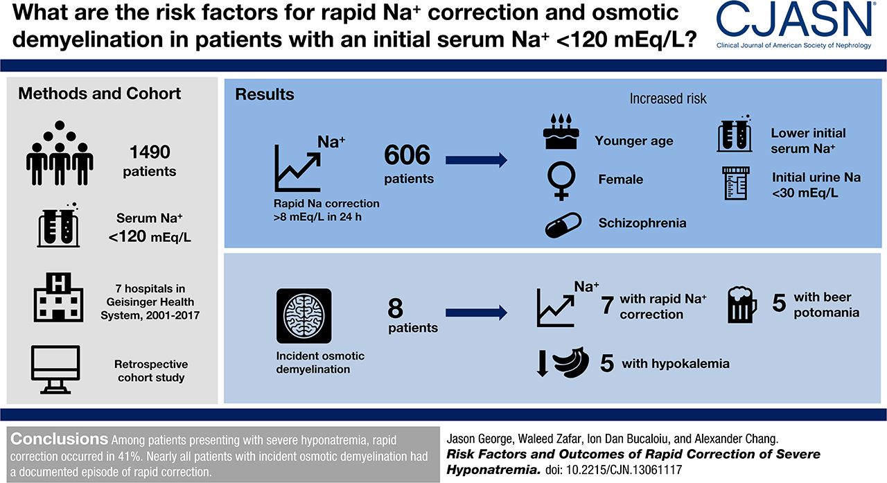
George, Zafar, Bucaloiu, and Chang analyzed the 7 hospitals in the Geisinger Health System to look at the incidence and risk factors for rapid sodium correction and osmotic demyelination.

They included adults with an initial sodium less than 120 mmol/L.

Their primary endpoint was an increase in sodium of more than 8 mmol/L in the first 24 hours.

Looking at the methods, it was not clear to me how they dealt with patients with multiple admissions for hyponatremia. Were they included multiple times? Did they only include the first admission?

They manually looked through every MRI report that was done after the index sodium to look for reports of ODS.

Results

They found 1,490 cases of severe hyponatremia with complete data that matched their inclusion criteria.

Median change in sodium in the first 24 hours was 6.8 mmol/L.

606 (41%) patients broke the speed limit of 8 mmol/L in the first 24 hours.

Risk factors for rapid correction

The authors provided a lot of depth on the risk factors for rapid correction. Here is the unadjusted risk factors.

Demographic:

  • Younger (average age of rapid correctors was 63 versus 68 in slow correctors)
  • Current smokers (40% of rapid correctors were smokers versus 26% of slow correctors)
  • Female gender

Medical history:

  • Depression (in 20% of rapid correctors versus 16% of slow correctors)
  • Schizophrenia (in 4% of rapid correctors versus 1% of slow correctors)
  • No history of hyponatremia (in 59% of rapid correctors versus 73% of slow correctors)
  • No history of chronic liver disease (in 6% of rapid correctors versus 8% of slow correctors)
  • No history of congestive heart failure (in 12% of rapid correctors versus 19% of slow correctors)
  • No history of cancer (in 19% of rapid correctors versus 25% of slow correctors)

Clinical data:

  • Lower body mass index (BMI of 26 in rapid correctors versus 28 in slow correctors)
  • Lower initial sodium (115 in rapid correctors vs 117 in slow correctors)
  • Lower initial urine sodium (35 in rapid correctors vs 43 in slow correctors)
  • Lower initial urine potassium (27 in rapid correctors vs 32 in slow correctors)
  • Lower initial urine osmolality (270 in rapid correctors vs 369 in slow correctors)
  • Have seizures (13% in rapid correctors versus 9% in slow correctors)

They used three different models for multivariate analysis to generate odds ratios for rapid correction of hyponatremia.

Some comments about the risk factors. I suspect the reason that depression actually represents SSRI induced SIADH that rapidly corrects when the drug is withdrawn.  The schizophrenia association may represent psychogenic polydipsia, a common manifestation of schizophrenia. Water restriction will result in prompt normalization of the sodium.

Liver, heart, and cancer are all associated with difficult to treat hyponatremia, so no surprise that they saw lower rates of rapid correction among people with those diseases.

The lower BMI and female gender association with rapid correction may be simply due to lower total body water making these patients more susceptible to rapid changes in serum sodium.

Lower urine sodium may indicate patients with volume depletion hyponatremia, a population predisposed to rapid correction of hyponatremia as soon as the volume depletion is corrected, releasing the hypothalamus from the volume stimulated release of ADH and resulting in rapid drop in ADH and rapid, uncontrolled correction of hyponatremia.

The lower urine osmolality likely represents patients with psychogenic polydipsia, tea and toast syndrome, or patients who are already in the midst of spontaneously correcting their sodiums, all patients who will rapidly correct their sodium.

The people who are seizing are a cohort with, presumably, acute symptomatic hyponatremia and rapid correction is less problematic.

Osmotic demyelination syndrome

There were 295 patients who had an MRI after having severe hyponatremia. Nine had radiologic evidence of osmotic demyelination. No patients had an ICD diagnosis of central pontine myelinolysis. One patient had MRI evidence of ODS prior to correction of the sodium. Consistent with medical consensus, nearly all of the patents with incident ODS had rapid correction of hyponatremia:

Of the eight (0.5%) patients who developed incident osmotic demyelination, seven (88%) had documented sodium correction > 8 mEq/L during any 24-hour period before brain MRI.

The one patient with evidence of ODS without rapid correction of hyponatremia had been admitted within a month with a sodium of 105. So it is possible the ODS occurred with that episode rather than the one captured in the study.

The authors listed the characteristics of the patients with ODS:

  • Hypovolemia in 75%
  • Beer potomania in 63%
  • Outpatient thiazide diuretic use in 25%
  • Alcohol use disorder 50%
  • Malnutrition in 50%
  • Hypokalemia in 63%

Having hypovolemia in 75% and beer potomania in 63% means that some patients had to have both of these diagnosis. This portrays a misunderstanding of what constitutes beer potomania. Beer potomania induced hyponatremia is not merely a patient who drinks beer and develops hyponatremia, instead it represents a patient who is unable to clear the water volume in beer due to inadequate solute intake. These patients have maximally dilute urine. On the other hand, patients with volume induced hyponatremia are unable to clear excess water because of hypovolemia-induced ADH release. In one case you have high ADH activity and concentrated urine (volume depletion) and in the other case you have suppression of ADH and dilute urine. There can not be a meaningful combination of the two disorders.

Alcohol use, malnutrition and hypokalemia are all previously recognized risk factors for ODS.

Three patients had received 3% saline for acute neurologic symptoms of hyponatremia. This is critical information because it shows that just because someone has acute manifestations of hyponatremia and an indication for 3%, one must still pay attention to the speed limit. Often acute neurologic symptoms of hyponatremia is thought to indicate a failure to have compensated for chronic hyponatremia and therefore protective against ODS. This is not the case.

The finding that I found most interesting was the clinical outcomes of the patients with ODS.

Five patients with documented osmotic demyelination had recovery with no neurologic deficits, two patients died from unrelated causes, and two were lost to follow-up.

They had 9 cases of ODS and they weren’t able to document even a single case of neurologic devastation. Five of the cases had documented neurologic recovery. This is how Burton Rose describes ODS:

And here is how Sarah and I described it in the fluids book

In my mind if you don’t spasmodic, mirthless laughter you don’t have ODS.

This is consistent with a previous study of dialysis patients with hyponatremia  who developed similarly clinically invisible ODS was diagnosed by MRI. From the discussion of Tarhan et al.

Contrary to its classic description, osmotic demyelination syndrome may develop without any presenting signs and symptoms in patients with end-stage renal disease who have undergone recent hemodialysis.

This largely asymptomatic ODS likely explains why not one of the 9 patients had an ICD diagnosis of central pontine myelinolysis (CPM). They did nt have any of the clinical manifestations of CPM.

When I read the abstract and saw that they had 9 cases of ODS in their cohort I jumed out of my shorts. This would be the largest series of DS ever. After reading the study I realize that actually we are in a new era of hyponatrmia where ODS can be devastating (read the three case reports in Brunner et al’s prospective study of MRI evaluation, PDF available on SciHub) or asymptomatic.

New hyponatremia data

In the last couple of months there has been an outpouring of new hyponatremia data and resources. The first I want to discuss is data on the speed of sodium correction with tolvaptan.

Juan Carlos lead a group who looked at the speed of sodium rise with tolvaptan. The primary endpoint was the change in sodium at 24-hours in patients given 15 mg of tolvaptan.

All patients had to have failed fluid restriction to be included in the analysis.

For the purpose of the study, SIADH was defined as:

  • serum sodium concentration ≤ 130 mEq/L
  • serum osmolality ≤ 280mOsm/kg
  • urine osmolality > 100 mOsm/ kg
  • urine sodium excretion > 20 mEq/L

CHF induced hyponatremia was defined as:

  • serum sodium concentration ≤ 130 mEq/L
  • serum osmolality ≤ 280mOsm/kg
  • echo- cardiographic evidence of systolic or diastolic dysfunction
  • urine sodium excretion < 20 mEq/L (if not on diuretics)

All patients had to start with a tolvaptan dose of 15 mg.

The only other concurrent therapy allowed was fluid restriction. Patients who subsequently were started on D5 water, diuretics or salt tablets, had their data censored at the point where the additional therapies were added.

Diuretics are listed as an exclusion criteria but the CHF group were allowed to use them (an exclusion to the exclusion criteria). This is not well described in the methods.

NO DIURETICS!
Except for the half of the cohort that has heart failure.

After restricting the patients by their pre-specified exclusion criteria they had 28 patients with SIADH and 39 with CHF.

Table 1.

Remember how the urine sodium is supposed to be low in heart failure. Take a look at the elevated level found in this study. Conclusion: diuretics work. Also take a look at the low Bun and low uric acid in the SIADH group. These are really helpful in my experience at differentiating the cause in tricky cases.

Tolvaptan was much more effective in SIADH with an average change sodium of 0.80 mmol/L/hr versus 0.17 mmol/L/hr in CHF

Sodium went up by more than 12 mEq/L in 25% of patients with SIADH and 3% of patients with CHF.

Using linear mixed-effects models to conduct multivariable repeated-measures analysis the investigators found:

  • In SIADH a lower serum sodium (<120 mmol/L) and lower serum urea (<6 mg/dL) were risk factors for rapid correction of sodium.
  • In CHF, only serum urea was a risk factor for rapid correction

This is what these variables look like when mixed together (data for SIADH patients)

The discussion includes this tidbit where the investigators try to explain why there is a more dramatic response in SIADH than in CHF.

As seen in Table 1, average kidney function of patients with SIADH was significantly greater than that of patients with CHF. As shown in Figures 4 and 5, a total of 8 of 39 patients with CHF and 1 of 28 patients with SIADH had serum creatinine concentrations > 1.5 mg/dL. Thus, difference in kidney function may account for the observed difference in therapeutic response between the SIADH and CHF groups.

I don’t find this argument convincing because kidney function was tested to see if it predicted response and though eGFR did correlate with response to tolvaptan in SIADH, it was not an independent predictor of response and was not a predictor of response at all in CHF.

In the SIADH cohort, age and baseline values for serum sodium, serum osmolality, SUN, serum creatinine, MDRD, and CKD-EPI significantly correlated with the magnitude of increase in serum sodium concentration during the first 24 hours of therapy. Unlike those parameters, no significant correlation was found between the initial 24-hour increase in serum sodium concentration and either body weight, body mass index, or baseline urine sodium, urine osmolality, serum uric acid, or serum potassium value. In the CHF cohort, baseline serum sodium, serum osmolality, SUN, serum creatinine, and serum potassium values significantly correlated with the 24-hour increase in serum sodium concentration. Conversely, no significant correlation was found between the initial 24-hour increase in serum sodium concentration and either age, body weight, body mass index, or baseline urine sodium, urine osmolality, MDRD, and CKD-EPI values.

This article is accompanied by an editorial by NephMadness Selection Committee member Richard Sterns. He does a nice job describing why this rapid increase in sodium in SIADH show in Morris’ paper was not also seen in Schrier’s SALT 1 and 2 paper. In that phase 3 trial that lead to the approval of tolvaptan, there were 51 patients patients with SIADH, and only 3 of them corrected too fast. This is 6%, well below the 25% found in Morris’ study. Sterns points out the relatively high sodiums found in SALT study (no one below 120 and only 30 had a sodium below 130) as a likely explanation.

Sterns wraps up his editorial with a neat description of the pharmacokinetics of tolvaptan and arguing for dosing the drug at 3.75 mg and then repeating the dose as needed every 6 hours to titrate the change in the sodium level.  Clever.

The minimally effective tolvaptan plasma concentration to increase urine output is approx. 25 ng/mL, and maximal increases in output occur when tolvaptan concentrations exceed 100 ng/mL. Levels > 25 ng/mL are achieved by doses as low as 3.75 mg, but do not remain at this level for long because the half-life for this dose is a little more than 4 hours. A 15-mg dose achieves peak plasma concentra- tions well above 100 ng/mL in patients with SIADH, enough to sustain a maximum water diuresis for more than 4 hours. A maximum water diuresis can increase the serum sodium concentration by >2.5 mEq/L per hour, yet it is not clear why this would be desired.

The standard practice in the United States is to administer 15 mg of tolvaptan and then encourage water intake to offset the resulting variable (and often large) water losses. Considering the high price of the drug in the United States (w$300 per tablet), this practice is basically flushing money down the toilet…

…A much more desirable outcome in patients with severe hyponatremia would be a modest but sustained increase in urine volume with a resulting slow steady increase in serum sodium concentration. If urine volumes were less massive, free-water restriction could be continued to avoid unwanted exacerbation of hypona- tremia. Theoretically, the desired response could be achieved with initial doses of 3.75 mg, repeating or increasing the dose every 6 hours if necessary, based on results of urine output and/or serum sodium levels measured before each dose, until the target increase in serum sodium level for the day is achieved.

Another source of additional insight on the study is an interview by Tim Yau of Juan Carlos at AJKDbog.org.

The Stanford short course on medical informatics, circa 1995.

After I graduated Medical School, I went to Stanford for a one week course on computer informatics. It was 1995. I had seen the world wide web before but this was my first exposure to HTML editing. We were shown expert diagnostics systems and an electronic medical record. It was Tomorrow Land for how the computer was going to shape the future of medicine.

The class was organized by Edward Shortliffe, At the time he was famous for this textbook:

It was published in 1990 and a number of our lectures came from chapters in this book. We think of the computerization of medicine as being a very contemporary subject, but Shortliffe was a co-author of a book titled “Readings in Medical Artificial Intelligence. The First Decade,” published in 1984!

The most important thing that I experienced in that course was Bayesian logic. There was a whole day on computer-aided diagnosis and as part of this, there was a lecture on the mathematics of pre-test and post test probability. Learning that there was a mathematical way to make sense of the uncertainty that had been a consistent companion on the wards was a revelation. I had travelled across the country to Palo Alto and Dr. Shortliffe had pulled the curtains of confusion from my eyes. He had shown me the science of medical decision making. It was a revelation.

At that time I was carrying around an HP200 lx, 1990’s ubercalculator/PDA.

It had an amazing programable calculator. I entered the equations for post-test probability and after class excitedly went up to Dr. Shortliffe and explained that I was sure that I could research the sensitivity and specificity of all the tests I needed, but I had never come across any data on the pre-test probability. I wanted to know where I could find that information. He looked at me and told me that the pre-test probability is your intuition as a doctor. You had to assign your own pre-test probability based on your history and physical and other pieces of data.

Intuition…

This detailed lecture with mathematical certainty was at its very core just human, fallible, intuition.

It crushed me. Math wasn’t going to save me.

I hadn’t thought of that moment in my medical education journey until I read The Laws of Medicine. Law 1: A strong intuition is much more powerful than a weak test.

Get a copy of this book and read it, so you can discuss it with #NephJC in the Summer Book Club this August.