Site improvement

If you have been annoyed at all of the broken links at PBFluids. My apologies. Blogger doesn’t host any files so to use them you need to host elsewhere. For this reason alone I recommend WordPress or SquareSpace for people who want to start blogging.

Tonight I fixed the Handouts tab. A few weeks ago I fixed the Books tab. All of those links now work. Next up: the Lectures tab.

Question: Should old lectures that may contain out of date material be taken down, or remain up?

Fuck you Wikipedia

The Wikipedia entry for Journal Club used to have a comprehensive list of Twitter-based Journal Clubs. It looked like this

I went to add another journal club to the list yesterday (hello Journal of Hospital Medicine and #JHMChat) and it was gone. I went into the history and discovered that Twitter Journal Clubs are not notable so on September 28th that list was taken from the world. Sorry.
I’m thinking of a new Wikipedia tag line, how does this sound: Wikipedia, where a comprehensive list of pornographic actresses belongs in the encyclopedia, but Twitter Journal Clubs? Not notable.

Using visual abstracts in presentations

This past Saturday I gave a talk at the ACP of Michigan on SGLT2 inhibitors.

The talk went well, except my HDMI to thunderbolt converter failed in a big way and I had to export the presentation to Powerpoint and run it off a Windows Machine. Yuck.

I used visual abstracts from EMPA-REG and CANVAS as a significant part of the presentation. This segment demonstrates how I used them. What killed me was the cool animation, where the third panel flips to reveal the renal outcomes, was handled with complete incompetence by PowerPoint. Otherwise PowerPoint did a pretty good job displaying my slide, but botching my favorite animation in the entire presentation is bordering on unforgivable.


SGLT2i renal outcomes from joel topf on Vimeo.

Here are the two visual abstracts in question:

This slide isn’t in the above video. I will eventually get the whole presentation up, but I love this one so much I had to share.

Speaking of sharing. This Wednesday, Dr. Christos Argyropoulos will kick off the first Tubular Talk with a presentation on SGLT2 inhibitors. Should be great check out all of the details at GlomCon.

Things I want to write about eventually: Exercise induced rhabdomyolysis

Exertional Rhabdomyolysis during a 246-km Continuous Running Race

SKENDERI, K. P., S. A. KAVOURAS, C. A. ANASTASIOU, N. YIANNAKOURIS and A. MATALAS. Exertional Rhabdomyolysis during a 246-km Continuous Running Race. Med. Sci. Sports Exerc., Vol. 38, No. 6, pp. 1054 – 1057, 2006. Background: To evaluate the effect of continuous, moderate-intensity ultraendurance running exercise on skeletal muscle and hepatic damage, as indicated by serum enzyme activity measured immediately following the race. Methods: Thirty-nine runners of the Spartathlon race (a 246-km continuous race from Athens to Sparta, Greece) who managed to complete the race within the 36-h limit participated in this study. Mean finishing time of the study participants was 33.3 T 0.5 h and their average age, height, and body mass were 41 T 1 yr, 174 T 1 cm, and 67.5 T 1.1 kg, respectively. Blood samples, taken a day before and immediately after completion of the race, were assayed for the following variables: creatine kinase (CK), lactate dehydrogenase (LDH), aspartate aminotransferase (AST), alanine aminotransferase (ALT), and gamma-glutamyltransferase (F-GT). Results: A dramatic increase in most of muscle and liver damage indicators was observed. The mean values for CK, LDH, AST, and ALT after the race were 43,763 T 6,764, 2,300 T 285, 1,182 T 165, and 264 T 37 IUILj1, respectively. These values were 29,384 T 4,327, 585 T 89, 5,615 T 902, and 1,606 T 331% higher than the corresponding values before the race (P G 0.001) for CK, LDH, AST, and ALT, respectively. However, there was not a significant increase in F-GT levels. Conclusion: Muscle and liver damage indicators were elevated at the highest level ever reported as a result of prolonged exercise, although no severe symptoms that required hospitalization were observed in any of the participants. The data suggest that even moderate-intensity exercise of prolonged duration can induce asymptomatic exertional rhabdomyolysis. Key Words: CREATINE KINASE, LACTATE DEHYDROGENASE, SPARTATHLON, ULTRAENDURANCE EXERCISE

So tasty.

Link

Get the NephRUN T-shirt While Fighting Multiple Myeloma

This tweet has taken off.

What do you call a nephrologists who likes to go jogging?

A Nephrun. pic.twitter.com/51pGZ0vQJh

— Joel Topf, MD FACP (@kidney_boy) October 3, 2017

A few people have expressed an interest in getting one for themselves. Okay, I’m a reasonable guy. How about this deal.

If you are going to Kidney Week and you donate $50 to the Multiple Myeloma Research Foundation for my trip to Everest I will get you a t-shirt. Your donation is even tax-deductible. This needs to happen in the next 10 days for me to get the shirts in time.

If you are not going to Kidney Week, donate $100 and I will send you a shirt.

Want a shirt and you already donated to the MMRF? Shoot me a tweet (DM or @) or e-mail and we’ll work this out.

After you have donated, fill out this form so I get you the right shirt and know how to get it delivered.

The shirt is really nice. The women’s version is a Hanes Ladies Cool Dri V-Neck Performance Shirt and the mens is the same shirt, but crew neck.

Have a nephrology question? #AskRenal to the rescue.

A couple of days ago, this came across my notifications

Prolonged OTC ingestion of sodium bicarbonate causing extremely low K+ (<1.5)
Any ideas as to the mechanism? @kidney_boy #askrenal

— Morgan (@Morgansb) September 27, 2017

The answer came quick.

How does bicarbonate cause hypokalemia

The video is here (complete with misspellings) and the Keynote file is here (with misspellings corrected)


How metabolic alkalosis causes hypokalemia from joel topf on Vimeo.

The beautiful #VisualAbstracts of the NEJM

This summer (I think) the NEJM began publishing visual abstracts on their twitter feed. Curiously, I was unable to find them on the page of the article that the visual abstracts references, or in the list of media types that you can search for.

The figure list on the right side does not include the striking visual abstract they created.

The “Browse Figures and Multimedia page has 19 different types of media, but visual abstract is not one of them.

The only way I could round up the visual abstracts was scrolling through the the NEJM Twitter feed. Here are the ones I found. Did I miss any?

Gorgeous work. Each one has a unique color palette and they have a pretty simple template, but three different ways of executing it. All of them look like they are from the same family except the tiotropium visual abstract. I really like that they give both the percentages and the raw numbers. No P-values or confidence intervals are found. These visual abstracts have as low an information density as I have seen. This is not a criticism, I think meh style has been increasing complexity to the detriment of my work. I need to turn up my inner NEJM.

Another question from OUWB

Hi Dr. Topf 

First of all, apologies for sending this via email but I do not have a Twitter account (I know, its the 21st century, who doesn’t?). 

I had a quick question regarding a practice problem I was doing. Rather than summarize the question for you, I included a screenshot so that you have the primary source with the explanation provided. Below, I also included my explanation for my reasoning for choosing that option. Basically, I am confused as to why the bicarb would be decreased in this scenario.

So the stem describes acute trauma. Specifically crush injuries, so you should be thinking rhabdomyolysis where the body gets turned inside out. In my very first lecture we talked about the intracellular atmosphere versus the extracellular atmosphere:

So expect increased potassium and phosphorus.
The vital signs show a patient with circulatory insufficiency, i.e. shock. There are some initial labs drawn from the blood and urine right before resuscitation is initiated. The urine shows an osmolality of 800 mmol/kg H2O (highly concentrated, indicating a lot of ADH activity) and a urine sodium of 5 mEq/L (very low, indicating increased activity of the renin angiotensin aldosterone system).
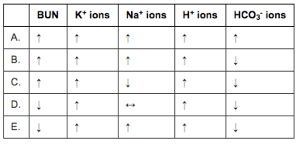
The question then asks you to predict the serum labs. Cool question. The best testing strategy here is to cross off ones that make no sense. Here are the foils:
BUN. This should be elevated as the patient moves to a pre-renal physiology. This leads to an increased filtration fraction to maintain GFR in the face of decreased renal plasma flow. This causes an increase in the osmolality in the efferent arteriole and vasa recta. this increases osmotic reabsorption of fluid from the proximal tubule. BUN flows passively with the fluid, decreasing renal urea clearance and increasing serum BUN. So D is wrong. E is wrong.
Potassium (K+) ions. All the choices show that it rises as the rhabdomyolysis from the crush injury releases loads of intracellular potassium. No answers are eliminated here.
Sodium (Na+) ions. We have a mishmash of choices here. This is difficult to predict. The increased ADH released due to shock would tend to lower the sodium. Increased aldosterone and renin would tend to increase the sodium. Since both are happening together I would expect them to balance each other out resulting in no change in sodium concentration. This is especially true since the stem specifically says there has not been much urine output I would go with D, for no change in sodium concentration. NOTE: About activation of the RAAS as a cause of hypernatremia. Hypernatremia is commonly listed as a symptom of Cushing syndrome and hyperaldosteronism so it is possible to have hypernatremia from (at least the pathological) activation of the renin angiotensin aldosterone system) but this is very unusual as increased in sodium concentration stimulates thirst which dilutes the sodium back to normal.
THIS COLUMN IS MESSED UP. THIS IS AN ERROR BY THE QUESTION AUTHOR.
Hydrogen (H+) ions. All the choices show that it rises. In shock we expect an increase in hydrogen ions as patients move to anaerobic metabolism due to inadequate perfusion (the functional definition of shock). No answers are eliminated here.
 
Bicarbonate (HCO3-) ions. Bicarbonate is the primary buffer in the body. Increases in hydrogen ions will be buffered by bicarbonate and bicarbonate will be consumed.
So the right answer is bicarbonate will be decreased. This eliminates answer A.
This leaves us with B or C and the question hangs on what the sodium will do and the reality is the change in sodium is unknowable. Shit question. Sorry.
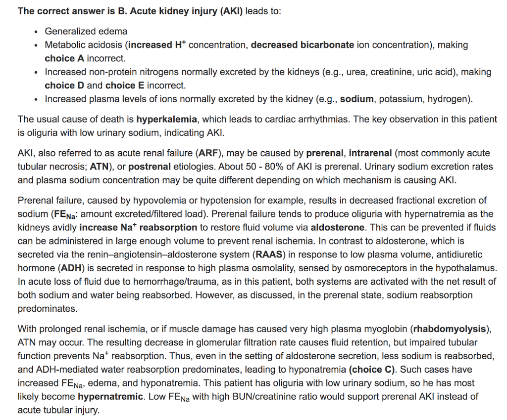
Here is what Kaplan claims to be true:
The forth bullet point is the one where the question fails. It is true that there is accumulation of plasma electrolytes in acute kidney injury. The problem is that these are electrolytes are measured as concentrations and there is also an accumulation of water (which is normally excreted by the kidney). This means that the effect on concentration is variable. Some, like hydrogen and potassium, reliably increase in AKI, but sodium is often decreased in AKI. Maybe the question writers were looking at an unconventional way measuring ions in the the plasma (as total amount rather than concentration).
The line in the answer that pre-renal azotemia is associated with hypernatremia is just wrong. You will encounter numerous patients with pre-renal disease that will have simultaneous hyponatremia. It is impossible to predict the serum sodium concentration from the volume status. This question reinforces the worst instincts of med students when it comes to predicting serum sodium. As I emphasized in the lecture, volume regulation and osmoregulation have two different regulatory systems and, though there is some cross talk, they are largely independent from each other.
The last paragraph tries to make the case that hyponatremia is more common in ATN while hypernatremia predominates in pre-azotemia. This is total fiction and does not exist. Though there can be more urinary sodium in ATN, if the patient is oliguric, it doesn’t matter how high the urine sodium concentration is, if the urine volume volume is close to zero there will not be much urinary sodium excretion.
Distinguishing between pre-renal azotemia and ATN is a constant problem in nephrology. Trust me you can’t make the determination by looking at the serum sodium. It aint that easy.
This question writer should never write another question about sodium.

 I posted this to twitter. The subsequent discussion was pretty interesting:

Kaplan Blows it on Hypernatremia and AKI

Medical student questions about nephrology

I have the honor of teaching at Oakland University William Beaumont School of Medicine. I teach sodium and water and acid-base to the second year medical students. After the lectures there is a steady stream of questions that start to fill my in-box. I answer the e-mail but I also post the questions and answers on PBFluids. Here is a directory to this year’s crop of Q&A.

Sodium concentration versus sodium content. With a second question on pseudohyponatremia vs false hyponatremia vs factitious hyponatremia

Macula densa and TG feedback. With a second question on whether SIADH is really euvolemic or just mostly euvolemic.

Breaking down an acid-base question.

More on euvolemic hyponatremia and how does this affect uric acid.

Urine chloride in non-anion gap metabolic acidosis, where does it come from?

Starling forces and GFR. With additional Q&A on edema, and metabolic acidosis and ammonia-genesis.