Hyperkalemia, medical management

The highest potassium I have ever seen is 9.9 mmol/L…

Don’t worry about that potassium of 9.9, the computer says it’s hemolyzed. twitter.com/kidney_boy/sta…
— Joel Topf (@kidney_boy) April 10, 2013

…however, it was a hemolyzed specimen so it is a tarnished victory. The patient is a dialysis patient in DKA and had a blood sugar of 925 at the time of the hyperkalemia.

I love that the only things not circled are a creatinine of 8.9 and BUN of 50.

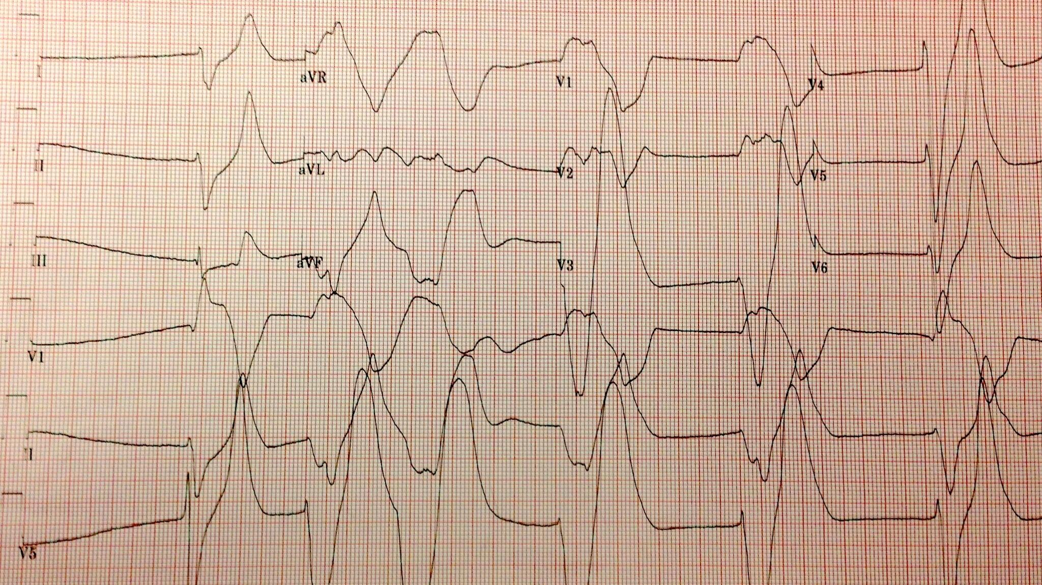
The patient was started on an insulin drip and one hour later his potassium was 5.2 mmol/L. A drop in the serum potassium of 4.7 mmol/L is profound and atypical. This is due to two factors:

  1. The initial serum potassium was not that high, despite the subtle EKG changes the real potassium had to be somewhat lower and falsely elevated due to the hemolysis.
  2. In DKA the hyperkalemia seen on presentation is due to a transcellular shift of potassium from the lack of insulin and increased extracellular osmolality (from the hyperglycemia), both of these are quickly reversible with IV insulin.
Adrogue et al created a formula to predict the potassium at admission in DKA.

K+= 25.4 – (3.02 x pH) + (0.001 x glucose) + (0.028 x Anion Gap)

The calculation is a totally failure in our patient: it comes to a predicted potassium of 5.6. Though the equation is not very predictive (R2= 0.25), the article does a nice job reviewing the mechanisms of hyperkalemia in DKA. This is a concept that is commonly misunderstood. Medical students are quick to rely on the mechanism of potassium moving out of cells in exchange for hydrogen moving into cells in the face of acidemia. Adrogue reviews the data that shows that this does not occur with the organic acids of DKA or lactic acidosis but that acidemia does inhibit the Na-K-ATPase.
Nice picture but this in not an important mechanism in the hyperkalemia of DKA.
The reason that acidemia does not generate hyperkalemia in DKA, is that the anions move into the cells along with the hydrogen ions, so there is no need to excrete potassium to maintain electroneutrality.
Horacio E Adrogue

Funny story about Adrogue, I was eating breakfast at Kidney Week in 2011 when I looked at the name tag of the guy sitting next to me, it was Horacio Adrogue! My chin hit the floor and I started to gush about how much I respected his work and how I loved his NEJM electrolyte reviews and how I was hoping he would autograph my chest and could I pick up his dry cleaning and… then he interrupted me to explain that he was not the Adrogue I was looking for. He was, in fact, The Man’s son and a transplant nephrologist of some regard. How humiliating. 

A nice review of the hyperkalemia and hyperglycemia in dialysis patients was published a couple of years ago by Tzamaloukas, a widely published investigator on the subject. I love this figure from the review which shows a curvilinear relationship of glucose and potassium in dialysis patients.

Nice graph except for micromoles of potassium
per mmol of glucose. Really? Could you make
it more obtuse?

One of the interesting conclusions that I learned from the review: one of the most important variables which affects how much the potassium will fall with insulin is the pre-treatments potassium level, the higher the potassium, the greater the response to insulin. The data from that conclusion  comes from this study: Serum potassium and acid-base parameters in severe dialysis-associated hyperglycemia treated with insulin therapy. It is an analysis of 43 episodes of hyperglycemia, half DKA and half non-ketotic hyperglycemia. Here is the money shot showing the relationship to initial potassium to drop in potassium:

What would you do for this patient? 

I will share the results in a week or so.

Addendum: some of the funnier tweets in response to my original tweet:

@kidney_boy probably artifact. Repeat ECG in am
— Lyle Shehane (@lyleshehane) April 10, 2013

@wanna_be_medic @kidney_boy It’s never good if your ECG looks like it was drawn by a five year old.
— Chump (@bungeechump) April 10, 2013

@kidney_boy unfortunately, it’s intravascular hemolysis.
— Michael Katz (@MGKatz036) April 10, 2013

@kidney_boy have patient follow up in Asystole Clinic in 3-5 days.
— GJ (@GregJNYC) April 11, 2013

We loved that comment so much we made it the Hyperkalemia Merit Badge:

But maybe we should have used:

Rules of Stone. Updated.

William J Stone is Chief of Nephrology at the Tennessee Valley VA and faculty at Vanderbilt Medical School. He discovered beta-2 microglobulin amyloidosis in long term dialysis patients. He describes the discovery here:

I discovered B2M amyloid in 2 hemodialysis patients during the late 1970’s and early 1980’s who sequentially broke both femoral necks. We were doing home hemodialysis on 75 VA patients, who lived an average of 220 miles away. One of them had a cystic knee lesion, which we biopsied. It was Congo red positive. A light bulb went off in my brain. All of the femoral neck tissue from both patients at joint replacement, misread as increased connective tissue, was full of amyloid. Workup for AA and AL was negative. The patient later died of lung cancer. At autopsy we scooped the amyloid out of a large humeral lesion. When sequenced in NYC, its subunit was intact B2M. To my knowledge, this has not been repeated.

In the early days of dialysis he was in Vietnam with Army and used dialysis for battlefield injuries, Stone again:

I was sent to the Third Field Hospital in Saigon from 1968-69, where we dialyzed battle casualties and falciparum malaria cases of AKI. I had completed a basic science nephrology fellowship at Cornell from 1965-67 and had never done dialysis before. We saved a lot of them using the old coil dialyzers.

One of his former fellows described him as, “One of those guys who can describe a case of almost anything you can imagine, across all of internal medicine.”

Dr. Stone has created the Rules of Stone, bits of wisdom that should guide doctors through the uncertainties of diagnosis and treatment.

Rules of Stone

  1. Anything can do anything. (WJS clarified: is for people who say things like a stroke alone cannot give you a high fever)
  2. Anything can do nothing. (WJS clarified: is for those doubters who say a patient on prednisone will have a tender abdomen if he has perforated an ulcer or a diverticulum)
  3. Nothing can do anything. (WJS clarified: refers to self-inflicted illness; e.g. IV injectors of dissolved pain pills can have multisystem disease)
  4. Nothing works every time.
  5. No lab or diagnostic test is perfect.
  6. No disease is always predictable.
  7. Just because you can do something doesn’t mean you should do something.
  8. A patient known to have x,y, and z doesn’t necessarily have x, y, and z.
  9. A person with an illness similar to a previous one probably has it again.
  10. No list is complete.
  11. The more drugs there are for a disease the less likely they are to benefit it.
  12. There are no uninteresting patients (just disinterested physicians).
  13. Four drugs are more toxic then two.
  14. Always guess 20% if you don’t know the real answer.
  15. No drug has been proven useless until it has been tried in scleroderma and ALS.
  16. Just because you failed to diagnose the cause of X in the past doesn’t mean you shouldn’t try again.
  17. Orneriness is best treated as an outpatient.
  18. If an older doctor writes an axiom or a diagram on a piece of paper, ask if you can have it.
  19. Common but unrelated diseases co-exist at least 1% of the time.
  20. If asked when you last played basketball, be able to answer with a day of the week.
  21. Before addressing non-compliance with diet and medicine #1, doctors add medicine #2.
  22. In a sick patient without a diagnosis, get invasive early.
  23. You cannot learn medicine at home, so the new residency hours rules make no sense. A cadre of doctors with inadequate experience is being created.
Two notes
  1. I am in total agreement with #23 
  2. The best safety data on creatine come from long term (negative) trials on using creatine in ALS, #15.

Pseudohyponatremia

Hyperglycemia causes pseudohyponatremia. The sodium is diluted by the osmotic movement of water from the intracellular to the extracellular compartment. I was taught the Katz conversion to correct the sodium, the sodium falls 1.6 mmol/L for every 100 mg/dL the glucose is over 100. This comes from purely theoretical work and was published in a letter the NEJM in 1972.

In the 1999 Hillier et al published empiric data that showed the ratio to be 2.4 rather than 1.6.

This ratio is now has been adopted by Mass General Handbook of Internal Medicine.

When ever I encounter hypernatremia I use both formulas and I consistently found that the Hillier estimated the final sodium. I wanted to do a study where I looked at hyperglycemia in dialysis patients and measured estimated final sodium versus actual final sodium to see which calculation worked better. It is a compelling study cohort because the lack of urine output guards against renal losses, a potential source of error. Well, the study has been done. Tzamaloukas et al, published a nice study of hyperglycemic dialysis patients and found the ratio of change in glucose to change in sodium was 100:1.5, almost exactly the same as Katz’s calculation and consistant with my experience.

The y-axis is the Katz estimate minus the actual final sodium. so a perfect estimate is zero. The x-axis is the average of the Katz and actual final sodium. The Katz conversion work well across a range of actual sodium levels.


Eat it Hillier.

Update, Dr. Rondon, in a comment below and Martijn vd Hoogen on Twitter, believe that I made a mistake calling hyperglycemic hyponatremia, an example of Pseudohyponatremia. There is some precedent for this position but it is not universal. See this editorial by the American Association for Clinical Chemistry, or McGraw Hill Concise Dictionary of Modern Medicine

We care about the serum sodium because of its effect on serum tonicity. When the serum sodium doesn’t represent the serum tonicity, the sodium is lying, I call that pseudohyponatremia. Pseudohyponatremia comes in two varieties: normal osmolality and high osmolality. The high version is what I am talking about in this post, the low version is what Drs. Rondon and Martijn vd Hoogen are referring to in there comments/tweets. More information on that can be found here.

@kidney_boy is this pseudohyponatremia? I thought this is dilutional hyponatremia, like mannitol. Pseudohyponatriemia is lab test error.
— Martijn vd Hoogen (@MWF_vd_Hoogen) April 14, 2013

iPhone urine micrographs

Calcium oxalate crystals
dirty granular casts indicative of ATN in a field of hematuria.
(The top left corner of the longer cast looks a little red cell cast-like)

Broad waxy cast, indicative of chronic kidney disease in a field of RBCs

New Nephrology merit badge for successfully using your iPhone to make a urine micrograph.

How do you determine the severity of SIADH? Updated with Video

There are a number of ways to grade the severity of SIADH. The most obvious is to look at how low the sodium is. The problem with this is that it largely depends on how much water a patient is drinking and is not solely dependent on the severity of the SIADH. A patient with mild SIADH that is started on hypotonic fluids will have a much lower sodium than a patient with severe hyponatremia who is adherent with her water restriction and urea tablets.

I assess the severity of SIADH by looking at the electrolyte content of the urine. Here is a doozy:

  • Urine sodium: 134
  • Urine Potassium: 62
  • Urine osmolality: 777
But this strategy suffers from a similar limitation, the urine electrolyte are affected by factors beyond the severity of the SIADH. Patients with SIADH are in sodium balance, that means that all the sodium they ingest is excreted. Increased sodium intake will be reflected by increased urine sodium, the same goes for dietary potassium and urinary potassium. The above labs came after the patient was given isotonic saline for 24-hours. The serum sodium fell from 128 to 117 with saline.
The true measure of SIADH severity probably is simply the urine osmolality.

Creatine is not just Creatinine misspelled

A few years ago I had a pre-med student shadow me on the dialysis service for a week or two. I had a hard time teaching him because he was so early in his medical education. I had him investigate an issue that he had some personal contact with and I think he did a nice job. The question was, “Is creatine nephrotoxic (bad for the kidneys).”

From this commercial sight, their
information is pretty tight

Creatine is synthesized by the body from the amino acids arginine, glycine, and methionine. It is phosphorylated by ATP to form phosphocreatine which is stored in muscle cells and acts as an instant energy source. When exercise depletes ATP, phosphocreatine rapidly restores ATP from ADP. Skeletal muscles typically have 3 times as much phosphocreatine as ATP. After being used up creatine is excreted as creatinine (and you thought creatinine’s purpose was to measure renal function).

The student, DJ_Scary, put together the following presentation on the biology and nephrotoxicity of creatine.

Medicine tends to be pretty puritanical. If it feels good, don’t do it. If it it feels bad, do it more. Eat your vegetables. Exercise every day. Don’t drink. Don’t smoke, Eat less red meat. Don’t put so much salt on your food. When it comes to performance enhancing drugs, the knee jerk and conservative response is the same. Avoid protein supplements. Don’t use anabolic steroids. Creatine will damage your kidneys. Some of this advice is wise, Joshua Schwimmer showed that anabolic steroids can cause FSGS (anybody ever test Zo or Sean Elliott for steroids?).

Creatine can double, triple or quadruple a patients serum creatinine. The math on how that works is shown in the video.
Note for the equations to be valid the following assumptions and units need to be used:

  • CrCl: ml/min
  • Creatinine: mg per 24 hours
  • Serum creatinine: mg/dL

The point of the video is that creatine will increase your creatinine and not affect your creatinine clearance or GFR which are the important variables. This is a situation where one cannot trust the estimated GFR formulas.

The medical puritan tells patients not to use creatinine because it can damage the kidney. This is not true. There is no data, beyond some pretty sketchy case reports that creatine can damage the kidney. Long term follow up with medical use of creatine shows no harm (randomized, placebo controlled data!). It seem convincingly safe (Oh, you wanted a Cochrane Meta analysis, we got that here). It will always be safer and more conservative for physicians to tell patients not to take a substance. (One year follow-up too short for you, how does 5 years of follow-up taste?) Patients deserve honest, unbiased answers about what different substances and behaviors do to their bodies and if physicians provide them with the same, old, predictable, puritan, advise, they will bypass doctors and we will lose our role in health advice.

To see a range of nephrologist views on creatine, take a look at the replies to this tweet.

Creatine: Bad for the kidneys or just bad for the kidney test (creatinine)?
— Joel Topf (@kidney_boy) March 5, 2013